Neurospora crassa genetic assay by fluorescence microscopy and the survey of mushrooms
Objetives:
1)Used fluorescence microscopy to identify the progeny of Neurospora, familiar with procedure of fluorescence microscope.
2)Familiar with the common mushroom in field.
Materials and Methods
1: N. crassa: SMRP10 x NCAC021-1.
2: Field-collected mushroom.
3: Water agar medium in Petri dishes.
4: A kit containing microscope slides, cover slips, needles, and transfer loops.
5: Immersion oil and dropper bottles for water to suspend specimens.
6: Lens paper for cleaning objectives, and Kim wipes for working with microscopic slides and specimens.
7: Bunsen burner, dissecting needle.
8: Microscopy: Canon PowerShot SD550 digital camera
Olympus SZ30 zoom stereo microscope Olympus CX31 compound microscope.
Olympus SZ30 zoom stereo microscope Olympus CX31 compound microscope.
Produces:
1: Pick out the perithecium from the culture, and gently crushing it on slide with a drop of dH2O, cover with cover slip.
2: Observe the sample under the fluorescence microscopy, take the pictures.
3: Survey the whole collected of mushrooms from the field. Take the pictures.
Observation:
I)
Fig.1 The cross culture
Fig. 2. Peritherium was observed from the culture
Fig. 3. Ascospore was released from the peritherium
Fig. 4. Ascospores was observed under the fluorescence microscopy
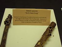
Fig.1. Nidula species
Fig.2. Cyathus alla
Fig. 3. Cariolus versicolor
Fig. 4. Daedoleopsis confragasa
Fig.5. Phallus species
Fig. 6. Xylaria species
Fig. 7. Morchella esculenta
Fig. 8. Laetiporus sulphureus
Fig. 9. Ganoderma lucidum
Discussion:
The progeny picked out from the perithecium is able to observe under the GFP fluorescence microscopy, indicating that the fluorescence protein was present in the progeny. Failed to find the asci that contain eight ascuspores, which theoretically half of them are able to observed the fluorescence and half of them are not. Probably I crushed too hard and destroyed all the intact asci.


No comments:
Post a Comment